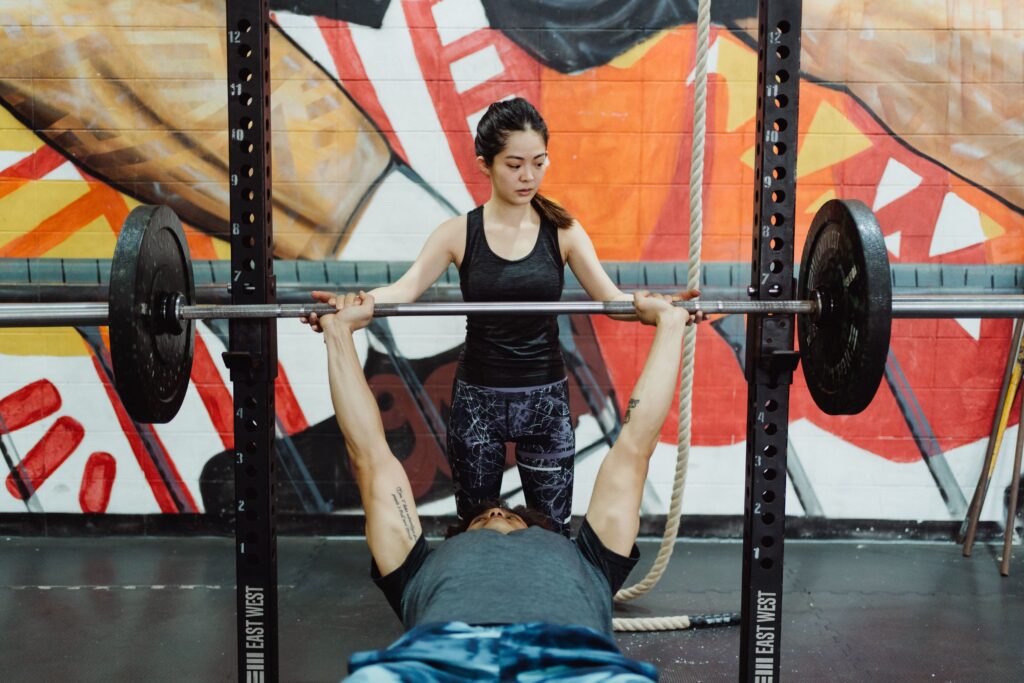
Fast reps vs slow reps, which is better for your gains?
Tempo in lifting is a hugely overlooked facet of lifting by many gymgoers and fitness lovers.
Jeremy Ethier explains everything you need to know about fast reps vs slow reps, and which is right for your goals.
Fast Reps vs Slow Reps – Which is Best for Maximum Muscle Growth?
“One often overlooked variable when it comes to training is lifting tempo – or how slow/fast you perform each repetition.”
Slow Reps
“In this video (see below) I’ll cover fast reps vs slow reps, and which one is better in terms of muscle growth. The main benefits of slow reps for mass is that it increases the time under tension throughout the set.”
Fast Reps
“On the other hand, the main benefit of using fast reps is that it enables you to use a heavier load or perform more reps when compared to slow reps. And although time under tension is reduced when fast reps are used, this actually doesn’t seem to hinder muscle growth.”
Fast Reps vs Slow Reps – What’s the Perfect Tempo?
“Thus, it’s clear that fast reps might be more beneficial for muscle growth. But how fast should you go and what’s the ideal rep speed?”
“Based on Schoenfeld’s meta-analysis, between 2 seconds to 6 seconds per rep seems optimal for muscle growth. But closer to 2 seconds (faster reps) seem to be slightly more beneficial.
Speed for the Concentric Portion of the Lift
“However, rather than obsessing over the best rep speed for muscle, a better approach would be to simply use a concentric speed that’s on the faster side but enables you to feel a strong mind-muscle connection.”
Speed for the Eccentric Portion of the Lift
“And for the eccentric portion of the lift, make sure you’re controlling the weight down as opposed to letting gravity do the work for you. Utilizing these two tips will help make the “ideal” lifting tempo easy to implement!”
Video – Fast Reps vs Slow Reps
Muscles of the Human Body
The muscles of the human body are the tissues that allow movement. Skeletal muscles make up about half of your body’s total weight, though they take up only about 20% of its mass.
Muscle tissue is categorized as either voluntary or involuntary.
Voluntary muscles are controlled by your brain and nervous system, and include those in your arms, legs and back.
Involuntary muscles control processes such as breathing, digestion and speech; they are also referred to as visceral (viscera) muscles or smooth muscle cells because they do not contain striations (stripes).
Fast Reps vs Slow Reps – Striated
These muscles comprise most voluntary skeletal muscle fibers found throughout the body except where large masses exist in the heart and large intestine walls which have fewer striations than typical skeletal muscle tissue due to low levels of physical activity associated with these areas
Fast Reps vs Slow Reps – Cardiac or Heart Muscle Cells
These cells allow contraction through electrical potentials created when sodium ions enter through ion channels at rest. Potassium ions exit through potassium channels allowing hyperpolarization between layers within each cell.
This happens along myofilaments which allows for contraction upon depolarization due to increased permeability between adjacent layers after binding together across gap junctions called syncytiums.
Head and Neck
The muscles of the head and neck are responsible for moving your face and head. They include a number of different muscles, including three that control eye movement:
- The levator palpebrae superioris elevates your eyelids to enable you to blink.
- The orbicularis oculi (a sack-like muscle) controls how much light enters your eye, as well as protecting them from bright light by squinting them closed.
- The rectus capitis posterior minor runs vertically up the back of your skull to help tilt it backwards and down when looking downward. It also helps rotate your neck from side-to-side or rotating it completely around so that you can look behind yourself (known as turning around).
Trapezius
Origin: Occipital bone, spinous process of C7-T12
Insertion: Lateral third of clavicle, acromion and spine of scapula
Action: Elevation of scapula (shoulder blade), depression and retraction of scapula(shoulder blade).
Splenius Capitis
The splenius capitis (pronounced: SPLEN-ee-us KAP-i-tis) is a small, flat muscle that lies along the side of your neck. It’s attached to several different bones in your spine and head, including your vertebrae, skull, scalene muscles and sternocleidomastoid muscles. The splenius capitis pulls together the upper portion of your neck and back so they move together in unison.
Sternocleidomastoid
- Sternocleidomastoid: The sternocleidomastoid is a paired muscle of the neck, which flexes the head to the same side and rotates it laterally. It is innervated by spinal nerves C1 through C3.
- Location: The sternocleidomastoid runs from the mastoid process behind your ear and attaches to your clavicle (collarbone). It lies deep to other muscles of your neck, and can be felt when you turn your chin toward your shoulder or tilt it up or down.
Temporalis
The temporalis is a muscle of mastication that originates from the temporal fossa and inserts on the coronoid process of the mandible. Temporalis is innervated by facial nerve.
Masseter
The masseter is a muscle of mastication and one of four muscles of the jaw. The other muscles are called temporalis, medial pterygoid, and lateral pterygoid. The masseter is the largest muscle in the human body. It originates from both sides of your jaw (temporalis) as well as from your zygomatic arch (zygomaticus major).
The facial nerve innervates both these areas independently so if you had damaged nerves due to trauma or surgery these movements would not be possible without proper rehabilitation therapy program designed specifically for individual needs.
Platysma
The platysma is a muscle of the human neck that is found between the skin and the superficial fascia. It is a superficial muscle, and is not visible from the surface. It covers most of the upper part of your chest area, where it attaches to a fibrous band called “platysma myoides” which lies along your front jawline. This muscle can be viewed in an anatomical dissection by pulling back on your skin at this point: you should feel two layers of tissue separate, revealing some red coloured tendon-like structures.
Digastricus
The digastricus is a muscle of mastication. It originates at the lateral half of the anterior surface of the body of the mandible and inserts into the anterior half of the medial surface of the hyoid bone, deep to its bony process. The digastricus acts to depress and protrude either side of your lower jaw (your chin).
The nerve supply for this muscle is from branches from three different nerves—the trigeminal nerve (cranial nerve V), facial nerve (cranial nerve VII), and hypoglossal nerve(cranial nerve XII).
Orbicularis Oculi
Orbicularis oculi is a muscle in the face that surrounds the eye socket. It helps close the eyelids and squints the eyes.
It also helps in chewing, especially when you chew your food with your mouth open (chewing with an open mouth requires more work for this muscle).
Zygomaticus Major and Minor
These two muscles are located on the cheekbone, and they work together to raise the corner of your mouth. The Zygomaticus Major is larger than its counterpart, but both muscles help you make expressions like smiling or laughing.
Conclusion
The human body is a complex machine, and it relies on many different muscles to function. Each muscle has its own role in the body and can be used for different purposes.
Use the information above to find the best lifting tempo for your workouts.
How to Increase Chest Size and Strength
Best Way to Train the Chest for Hypertrophy (Muscle Mass)